What is a hernia ?
In simple terms, a hernia can be explained as a defect in the walls of a container through which the contents inside the container come out, especially when pressure inside increases.
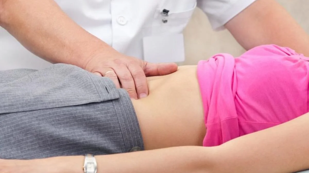
Hernias occur commonly in the abdominal (tummy and groin) region. Our abdomen is also like a container with its walls made up of muscles and fascia (tough fibrous tissue). The inside of the abdomen has many organs like small and large intestines, a pad of fat called omentum, liver, kidneys, spleen, etc. If there is a defect in the abdominal wall, the contents inside (most commonly the small intestine, which is very long and mobile, and the omental fat pad) tend to come out of the defect. This causes a bulge beneath the skin in that area. When the person lies down or pushes the contents, they go back inside the abdomen and the hernia disappears (reduces).
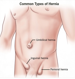
When the hernial defect occurs in the umbilical region it results in Umbilical Hernia.
When the hernial defect occurs in the groin area, it results in Inguinal or Femoral hernia.
Abdominal hernia may also occur at the site of a previous surgery. If the incision from the surgery has not healed well, it may result in a defect in the abdominal wall resulting in a hernia. Such a hernia is known as Incisional hernia.
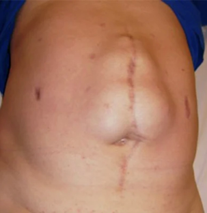
The above picture shows a hernia in relation to a scar from previous abdominal surgery .
What causes a hernia ?
Hernias can be present at birth (umbilical, inguinal, or groin hernias). They are known as congenital hernias. Most hernias that are seen in adults result from a combination of factors.
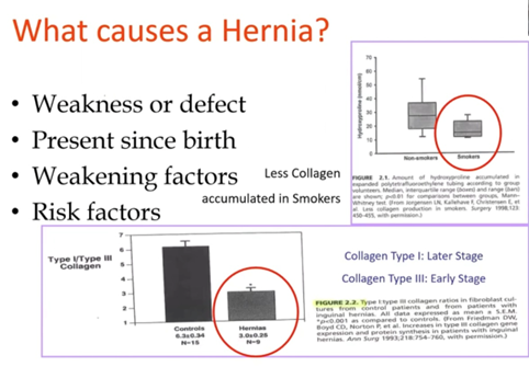
The building blocks of connective tissue (muscles, tendons, fascia, bones, etc.) in the human body is collagen. It has been seen that if a person has an increased amount of a certain type of collagen (viz type 3), they are prone to develop hernias.
Additional factors that result in increased stress and tears in muscles resulting in defects (hernias) are lifting heavy weights, severe chronic cough, severe constipation, etc.
Elderly sick patients with weak muscles too have a tendency to develop hernias.
What is the treatment for hernia ?
Since a hernia results from a physical defect in muscles and/or fascia, it needs to be physically closed by surgery. Medical treatment is not possible or effective in its treatment.
There are different surgical procedures for repair of hernias.
Open repair of hernia
This is a traditional method of repair. It involves a skin incision directly over or in close proximity to the hernia. The hernial contents are then reduced, and the defect is closed by using sutures or a mesh or both. A mesh is very commonly used nowadays to bolster the strength of a hernia repair to reduce the chances of recurrence. This procedure can be carried out under regional (spinal) anaesthesia and does not require general anaesthesia in most cases.
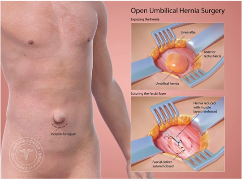
The above picture shows an open repair of an umbilical hernia. A skin incision is given just below or above the umbilicus, the protruding hernial sac with contents is reduced, and the defect repaired with sutures. Very often this is reinforced by application and fixation of a mesh.
Laparoscopic repair of hernia
This involves use of minimal access techniques. The area of the hernial defect is reached using very small cuts through which special instruments (a laparoscope - which transmits light and carries back magnified images onto a monitor via a camera attached to it and instruments like scissors, dissectors, etc.) are introduced. The contents of the hernia are reduced, and the defect is repaired by sutures and mesh.
The advantages of Laparoscopic repair are :
Early recovery
Less pain
Smaller scars - more cosmetic

Less infections
The above picture shows the introduction of small caliber trocars ( hollow cylinders) into the abdomen thru which laparoscopic surgical instruments like scissors, dissectors etc are inserted to perform surgical procedures.
Robotic assisted repair of hernias
Robotic assisted repairs have increasingly become more popular and feasible. These surgeries too use the minimal access techniques. It involves the use of robotic arms and instruments to perform surgery under complete control of the surgeon. The instruments reach the surgical area through minimal access (very small cuts).
Robotic assisted surgery has all the advantages of Laparoscopic surgery viz:
Early recovery
Less pain
Smaller scars - more cosmetic
Less infections
The added advantages of robotic assisted repair of hernias are :
3D visualisation of the operative field
Very precise dissection due to the increased freedom of movement of robotic instruments
Lesser pain as the movement at the entry point of instruments into the body is minimal in robotic surgery
Better ergonomics ( position during performing surgery) for the surgeon helps in surgeries with longer duration.
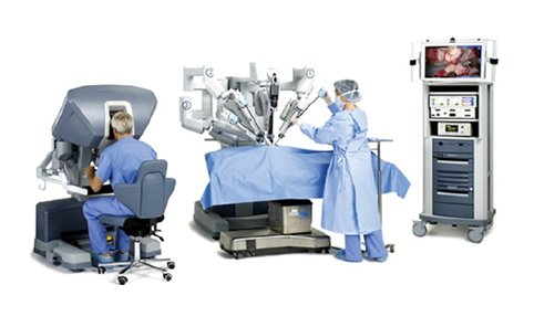
The above picture shows the arrangement for robotic surgery. The surgeon is sitting on a console from which he is able to get a 3-dimensional view of the surgical field and is able to move the instruments and camera with his hand controls. The middle area shows the patient on the table with the robotic arms carrying the instruments into the abdomen through small cuts. A surgical assistant is by the side. The right side of the picture shows a tower with a light source, an insufflator for the introduction of CO2 gas inside the abdomen, and instruments for diathermy cautery. On top of the tower is a monitor where the assistant and nursing staff can see the surgical field.
The above picture shows the arrangement for robotic surgery. The surgeon is sitting on a console from which he is able to get a 3-dimensional view of the surgical field and is able to move the instruments and camera with his hand controls. The middle area shows the patient on the table with the robotic arms carrying the instruments into the abdomen through small cuts. A surgical assistant is by the side. The right side of the picture shows a tower with a light source, an insufflator for the introduction of CO2 gas inside the abdomen, and instruments for diathermy cautery. On top of the tower is a monitor where the assistant and nursing staff can see the surgical field.
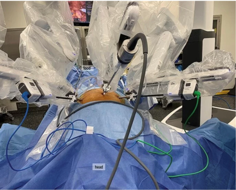
A view of the robotic arms with the instruments introduced inside the abdomen. The robotic arms are totally under control of the surgeon. They move only when they get signals from the hand movements of a surgeon.
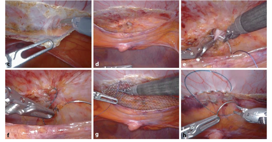
The picture below shows the inside view of Robotic assisted repair of Umbilical Hernia. Serial images show the dissection of flap and reduction of hernia sac followed by suture repair of the hernial defect and the reinforcement of the repair using a mesh. Robotic instruments are used for the surgery which results in very precise dissection and less trauma to tissues.